

Refine and Redefine Intervention Using High-Definition (Hi-Def) Technology
Andrew Kuhls-Gilcrist, PhD, DABR, Mark Hohn, Dale Marek, RT(R), Yiemeng Hoi, PhD
Medical Affairs, Interventional X-ray, Canon Medical Systems USA, Inc.
Swetadri Vasan Setlur Nagesh, PhD, Stephen Rudin, PhD, FAAPM,
Adnan Siddiqui, MD, PhD, FACS, FAHA, FAANS
Canon Stroke and Vascular Research Center University at Buffalo, Buffalo, NY
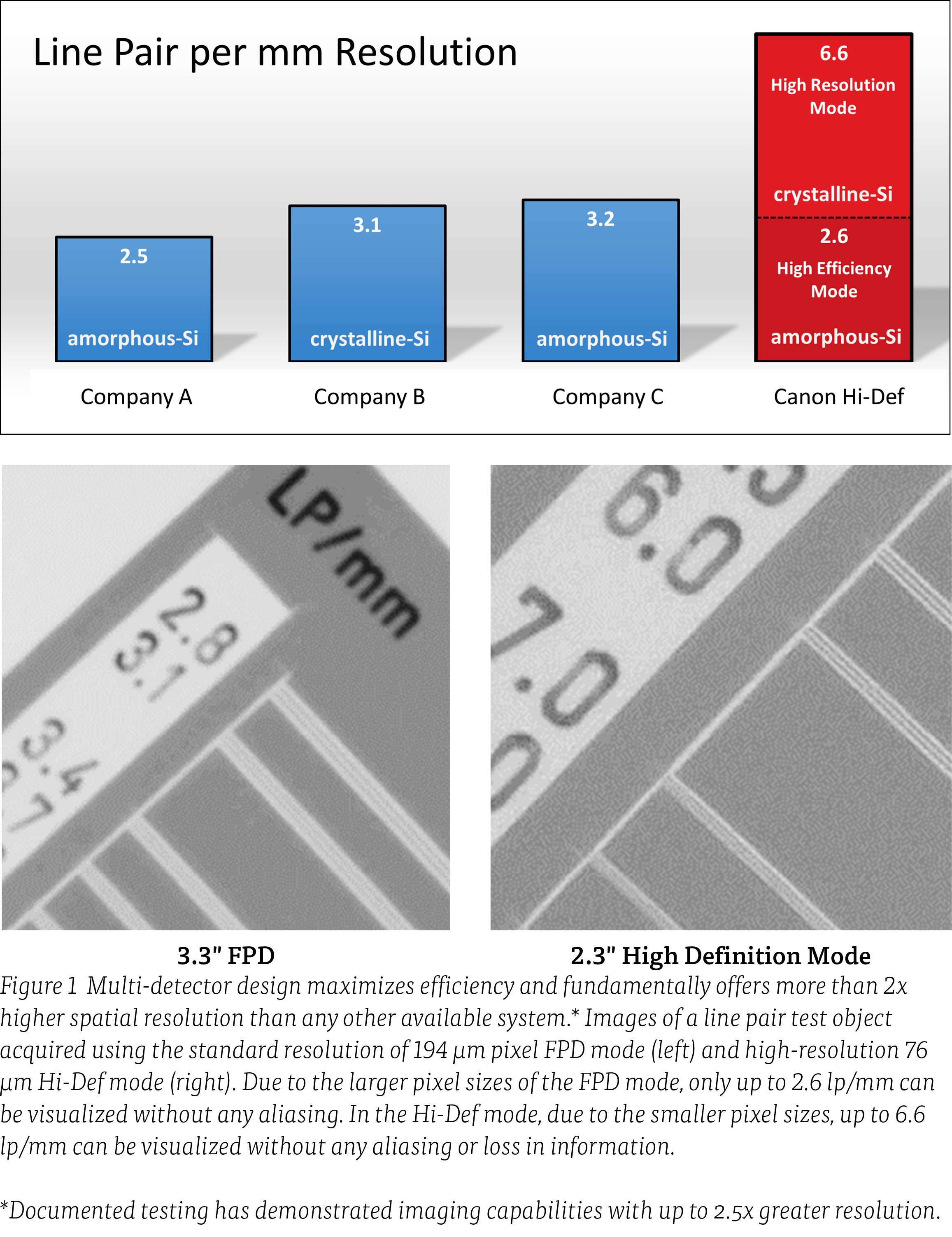
Modes Up to 2.5x Higher Spatial Resolution
Advancing minimally invasive treatments for neurovascular diseases such as stroke and aneurysms, places increasing demands for high definition real time imaging for aiding the interventionist in guiding the catheters upon identifying disease area, and to deploy treatment devices such as balloons, coils, stents, or flow diverters. Higher spatial resolution modes of the Hi-Def detector can provide sharper and visually improved images, as quantified using standard physical metrics, compared with those of traditional FPD images.1 For example, the modulation transfer functions (MTFs) of the new detector under Hi-Def mode and the FPD mode demonstrate the ability of the Hi-Def magnification modes to image spatial frequencies that would otherwise be aliased (i.e. not visualized) by the FPD magnification modes. Inevitably, Hi-Def outperforms the FPD at all spatial frequencies.5
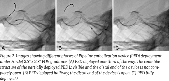
“The Hi-Def mode of the new detector system is equivalent to a microscope that can be used during critical stages of the intervention.”
— Adnan Siddiqui, MD, PhD, FACS, FAHA FAANS
Kaleida Health-Gates Vascular Institute, Buffalo, NY
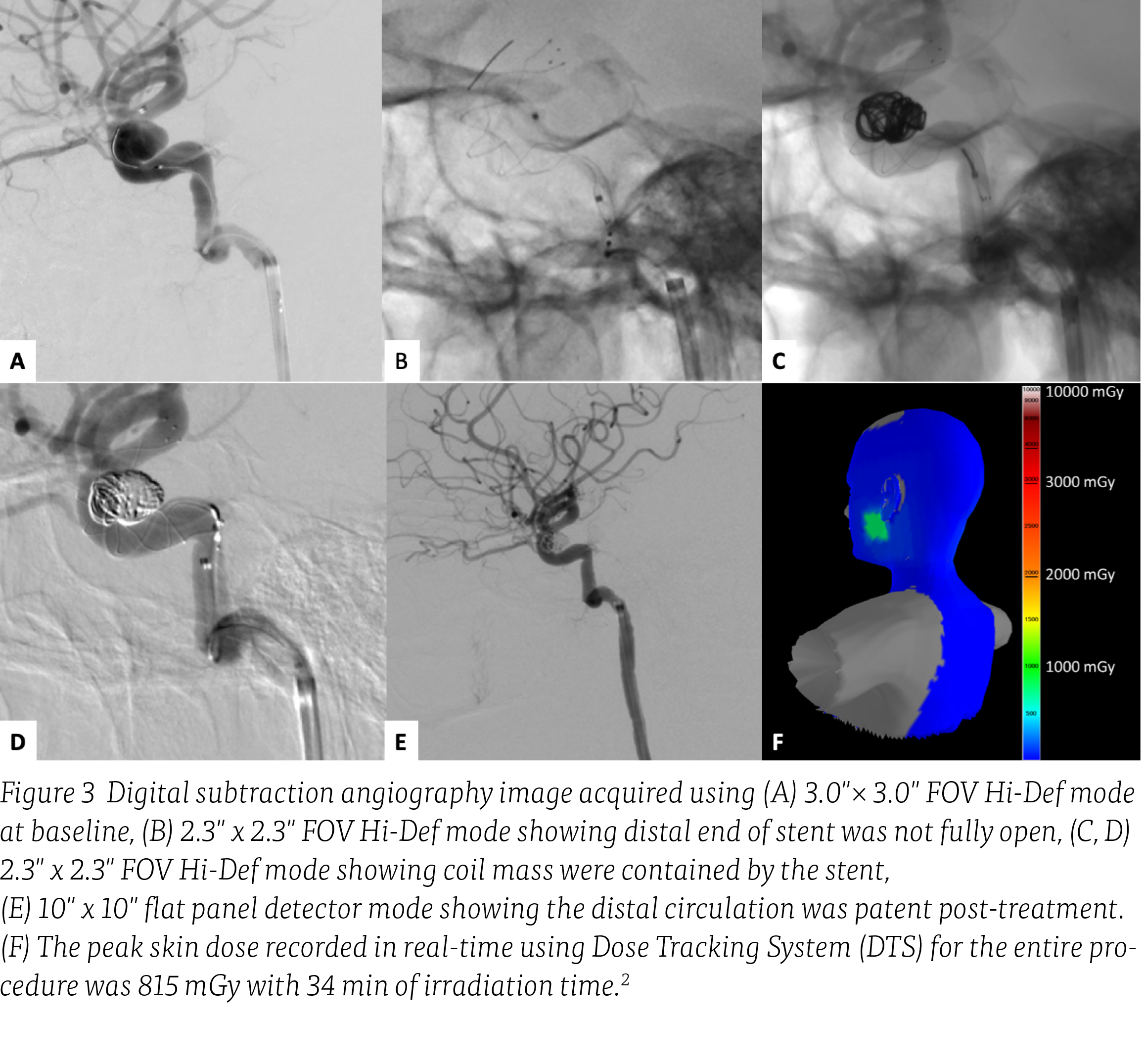
Case illustration2
A patient with no remarkable medical history presented to the emergency room with the worst headache of her life. Magnetic resonance angiography demonstrated a right carotid cavernous aneurysm measuring 11 × 7 mm. Lumbar puncture and head CT scan were negative for subarachnoid hemorrhage. The aneurysm size and location were confirmed with diagnostic cerebral angiography.
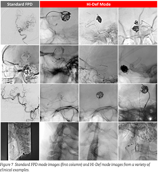
Under 2.3" x 2.3" FOV Hi-Def mode roadmap guidance, a 2.5Fr microcatheter over a 0.014" microwire was introduced and placed in the right distal M1 segment (Figure 3). An 2.4Fr microcatheter with a 45° angle was introduced concomitantly over the 0.014" microwire to cannulate the aneurysm neck. Under Hi-Def magnification, a single 9 × 33 mm coil was partially deployed into the aneurysm dome. Subsequently, a 4.5 × 23 mm LVIS Blue stent was introduced into the 2.5Fr microcatheter and deployment was started just distal to the aneurysm neck. Under 2.3" x 2.3" FOV Hi-Def mode, deployment showed good wall apposition throughout the curve of the cavernous carotid artery. The partially deployed coil was then fully deployed, followed by a second 5 × 20 mm coil. After the second coil deployment, DSA contrast runs were obtained and showed stasis in the aneurysm dome. The imaging mode was switched to large FOV, and a DSA contrast run was obtained to make sure the distal circulation was patent post-treatment. The patient was extubated and remained neurologically intact.
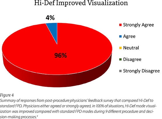
Physicians either agreed or strongly agreed, in 100% of situations, Hi-Def mode visualization was improved compared with standard FPD modes during the procedure and decision-making.
| Average Value | Pre Hi-Def Era | Post Hi-Def Era | % Diff | p-value |
|---|---|---|---|---|
| Procedure time (min) | 43.15 | 39.18 | -9.2% | < 0.001 |
| Cumulative Air Kerma (Gy) | 1.11 | 1.00 | -9.6% | 0.003 |
| Fluoro Time (min) | 19.12 | 17.48 | -8.6% | 0.004 |
| # of Pulses | 17,643 | 16,192 | -8.2% | 0.005 |
| # of 3D Spins | 0.30 | 0.26 | -12% | 0.023 |
| # of Irradiation events | 85.0 | 82.2 | -3.3% | 0.059 |
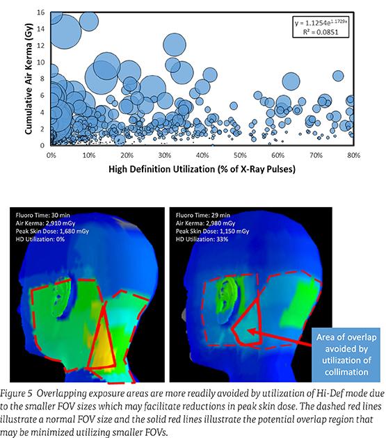
Figure 6 With results from over 6,300 neurointerventional procedures highlighting reduced procedure time, cumulative air kerma, fluoro time, and number of X-ray pulses. There was no correlation observed between high definition utilization and cumulative air kerma (top).
The bubble size relates to the total number of irradiation events. With results from over 6,300 neurointerventional procedures highlighting reduced procedure time, cumulative air kerma, fluoro time, and number of X-ray pulses (bottom).
Average procedure time, average fluoro time, the total number of X-ray pulses and cumulative air kerma were all significantly reduced by 8% or more.
The Best of Both Worlds:
Consistent Low Dose Performance
with Additional Hi-Def Modes
Consistent Low Dose Performance
with Additional Hi-Def Modes
The ability of Canon Medical’s Hi-Def technology to provide substantially increased spatial resolution modes can support complex endovascular procedures as clinical methods accessing smaller and more delicate structures are advanced and therapy moves towards more individualized, patient specific treatment. Hi-Def technology and increased visualization capabilities may well be fundamental in this next-generation era offering meaningful improvements to patient care. //
References
1. Setlur Nagesh SV, et al., Proc. SPIE 10573, Medical Imaging 2018: Physics of Medical Imaging, 1057365 (9 March 2018).
2. Setlur Nagesh SV, et al., J Neuroimaging. 2019 Sep;29(5):565-572.
3. Setlur Nagesh SV, et al., AJNR Am J Neuroradiol. 2019 Feb;40(2):302-308.
4. Setlur Nagesh SV, et al., J Neurointerv Surg. 2020 Sep;12(9):897-901.
5. Krebs J, et al., Proc. SPIE 10948, Medical Imaging 2019: Physics of Medical Imaging, 109480E (1 March 2019).
6. Miller DL, et al., J Vasc Interv Radiol. 2003 Jun;14(6):711-27.
Social Media


