

Initial Global Post Market Experience with PIQE



To the clinical MRI community, Precise IQ Engine (PIQE) offers the ability to generate images with higher in-plane matrix sizes from lower resolution images, often making this possible even at a faster acquisition time. Based on the advanced scientific principles of deep learning-based denoising and an image up-sampling process, PIQE can triple the in-plane acquired matrix size in both directions, producing higher image sharpness. With PIQE, valuable SNR is not traded for resolution but preserved and even enhanced due to denoising. More technical details explaining how PIQE works can be accessed by clicking the following link. Additionally, related articles including the validation process can be accessed in the "More Information" section at the bottom of this page.
As an integral component of the product release process and overall life cycle, it is especially important to monitor clinical adoption of new and innovative applications at the earliest stage. In partnership with the Johns Hopkins University (JHU) School of Medicine in Baltimore, Maryland, USA, and the Diagnostic Imaging Unit at Clinica Creu Blanca, Barcelona, Spain, Canon Medical Systems has provided the unique opportunity to evaluate product implementation immediately after initial release to the market. This provides an opportunity to confirm the outcomes of prior validation studies as well as expand our knowledge of clinical benefits and potential limitations. Both aspects are useful in building upon education, training and enabling a more seamless clinical adoption.
During this early post market implementation of PIQE, strategies involved the acquisition and multiple reconstructions of repeated scans with low and high acquisition matrices compared to the standard clinical protocol. At JHU, investigators sought to determine the lower limit on the acquired matrix size which could be reconstructed to produce a high-quality, high-resolution image with PIQE. Similarly, Crue Blanca compared their standard, already high-quality clinical protocols to an ultra-high-resolution acquisition and a faster acquisition acquired at a lower resolution and reconstructed to the same ultra-high resolution. Sample comparisons of image quality, resolutions and acquisition times are shown in Figures 1 - 5.
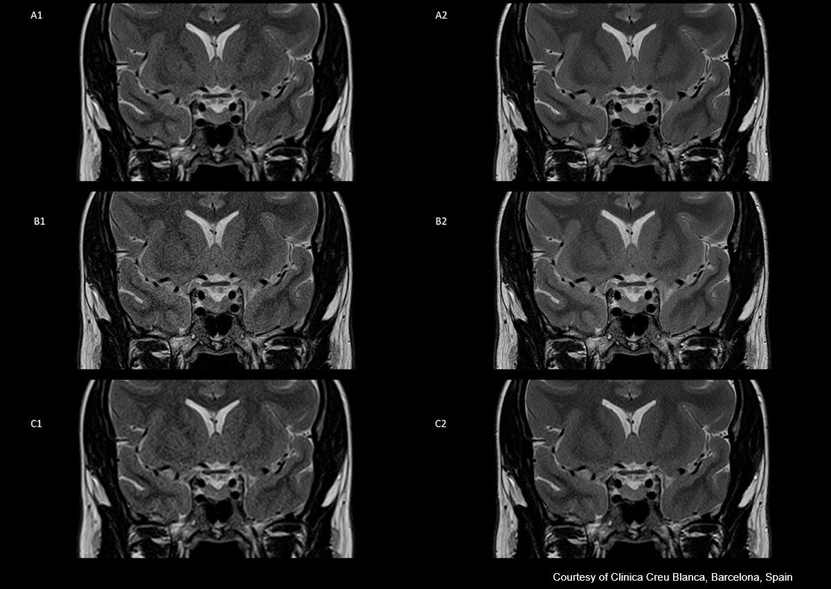
| Standard (A1) | Standard PIQE (A2) | High Resolution (B1) | High Resolution PIQE (B2) | Faster (C1) | Faster PIQE (C2) | |
| Acquired Matrix | 224x224 | 224x224 | 352x352 | 352x352 | 192x192 | 192x192 |
| Field of View (mm) | 150 | 150 | 170 | 170 | 150 | 150 |
| Slice Thickness (mm) | 2 | 2 | 2.2 | 2.2 | 2 | 2 |
| Scan Time | 2 min 12 sec | 2 min 12 sec | 2 min 25 sec | 2 min 25 sec | 1 min 5 sec | 1 min 5 sec |
| Reconstructed Matrix | 448x448 | 672x672 | 704x704 | 1056x1056 | 384x384 | 576x576 |
| Pixel spacing (mm) | 0.33 | 0.22 | 0.24 | 0.16 | 0.39 | 0.26 |
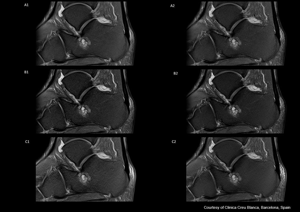
| Standard (A1) | Standard PIQE (A2) | High Resolution (B1) | High Resolution PIQE (B2) | Faster (C1) | Faster PIQE (C2) | |
| Acquired Matrix | 256x256 | 256x256 | 256x288 | 256x288 | 192x192 | |
| Field of View (mm) | 130 | 130 | 130 | 130 | 130 | 130 |
| Slice Thickness (mm) | 3 | 3 | 1.8 | 1.8 | 3 | 3 |
| Scan Time | 1 min 55 sec | 1 min 55 sec | 3 min 4 sec | 3 min 4 sec | 56 sec | 56 sec |
| Reconstructed Matrix | 512x512 | 768x768 | 576x576 | 864x864 | 384x384 | 576x576 |
| Pixel spacing (mm) | 0.25 | 0.17 | 0.23 | 0.15 | 0.34 | 0.23 |
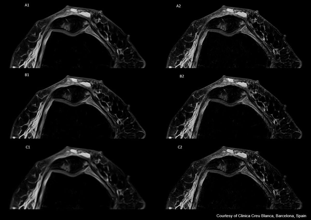
| Standard (A1) | Standard PIQE (A2) | High Resolution (B1) | High Resolution PIQE (B2) | Faster (C1) | Faster PIQE (C2) | |
| Acquired Matrix | 256x256 | 256x256 | 352x288 | 352x288 | 192x192 | 192x192 |
| Field of View (mm) | 160 | 160 | 160 | 160 | 160 | 160 |
| Slice Thickness (mm) | 2.5 | 2.5 | 2.2 | 2.2 | 2.5 | 2.5 |
| Scan Time | 2 min 11 sec | 2 min 11 sec | 3 min 19 sec | 3 min 19 sec | 56 sec | 56 sec |
| Reconstructed Matrix | 512x512 | 768x768 | 704x704 | 1056x1056 | 384x384 | 576x576 |
| Pixel spacing (mm) | 0.31 | 0.21 | 0.23 | 0.15 | 0.42 | 0.28 |
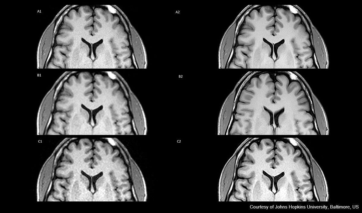
| Standard (A1) | Standard PIQE (A2) | Faster (B1) | Faster PIQE (B2) | Fast/Thin Slice (C1) | Fast/Thin Slice PIQE (C2) | |
| Acquired Matrix | 288x288 | 288x288 | 192x192 | 192x192 | 192x192 | 192x192 |
| Field of View (mm) | 230 | 230 | 230 | 230 | 230 | 230 |
| Slice Thickness (mm) | 4 | 4 | 4 | 4 | 2 | 2 |
| Scan Time | 2 min 44 sec | 2 min 44 sec | 1 min 18 sec | 1 min 18 sec | 2 min 36 sec | 2 min 36 sec |
| Reconstructed Matrix | 576x576 | 864x864 | 384x384 | 576x576 | 384x384 | 576x576 |
| Pixel spacing (mm) | 0.4 | 0.27 | 0.6 | 0.4 | 0.6 | 0.4 |
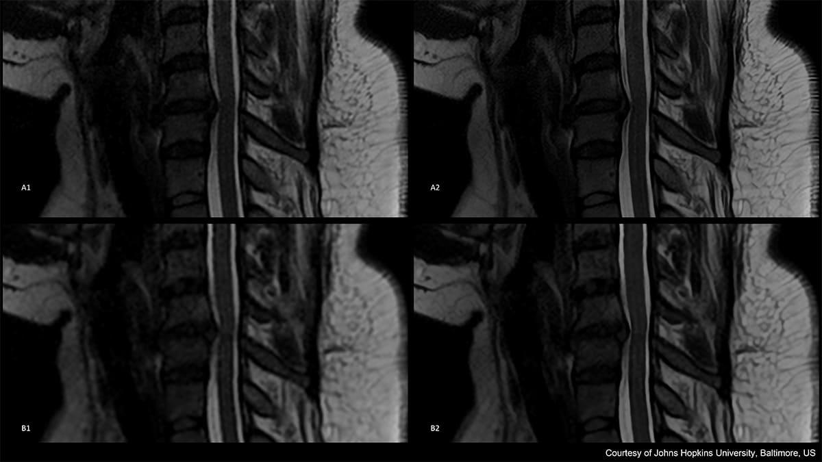
| Standard (A1) | Standard PIQE (A2) | Fast/Thin Slice (B1) | Fast/Thin Slice PIQE (B2) | |
| Acquired Matrix | 288x288 | 288x288 | 192x192 | 192x192 |
| Field of View (mm) | 200 | 200 | 200 | 200 |
| Slice Thickness (mm) | 3 | 3 | 2 | 2 |
| Scan Time | 2 min 29 sec | 2 min 29 sec | 2 min 39 sec | 2 min 39 sec |
| Reconstructed Matrix | 576x576 | 864x864 | 384x384 | 576x576 |
| Pixel spacing (mm) | 0.35 | 0.23 | 0.52 | 0.35 |
In summary, these initial experiences with PIQE on Vantage Orian (1.5T) and Vantage Galan 3T scanners have begun to showcase the unique value PIQE offers in overcoming MRI specific challenges. The traditional challenge of consistently and reliably achieving high image quality in a shortened acquisition time is no longer a big hurdle in MRI. Reconstruction with PIQE enables shortened acquisition times by lowering the acquired pixel resolution, while deep learning and up-sampling allows for the reconstruction of superior high-resolution images, preserving structural detail and removing the noise. PIQE can be utilized differently in a variety of clinical scenarios. When faster scan times are needed to improve patient comfort or when higher resolution images and/or additional thinner slices are needed to visualize super fine detailed structures, PIQE can offer a solution.
Avis de non-responsabilité
Certaines fonctionnalités présentées dans cette publication peuvent ne pas être disponibles commercialement sur tous les systèmes présentés ou peuvent nécessiter l'achat d'options supplémentaires. En raison des réglementations locales, certaines fonctionnalités commerciales incluses dans cette publication peuvent ne pas être disponibles dans certains pays. Veuillez contacter le représentant Canon Systèmes médicaux de votre région pour obtenir plus de détails et les informations les plus récentes.
La technologie d'apprentissage profond est utilisée dans la phase de conception du traitement de la reconstruction d'images. Le système lui-même n'a pas de capacités d'autoapprentissage. Le contenu de ce rapport comprend les opinions des auteurs basées sur leur expérience et leurs connaissances cliniques.
For more information

Inscription aux dernières informations sur l’IRM de Canon
Inscrivez-vous dès maintenant pour obtenir nos nouvelles publications et des mises à jour sur nos produits.*
Required information

Social Media


