

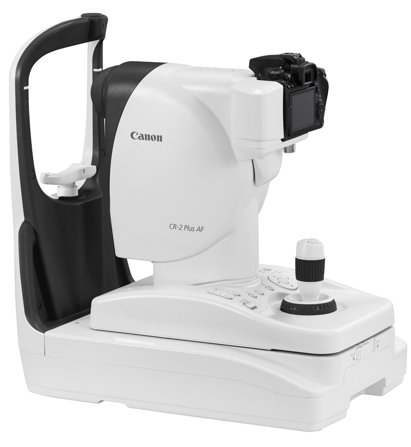
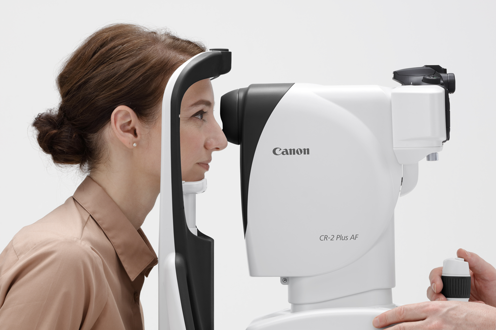
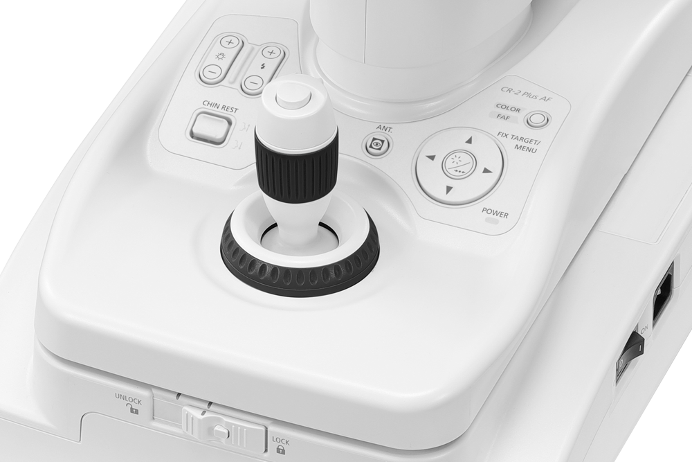
CR-2 Plus AF
See the smallest details, don’t miss pathology
Canon was the very first company to introduce a Non-Mydriatic Retinal Camera as far back as 1976.
Built on this legacy, the CR-2 Plus AF is equipped with superb Canon optics and Canon’s own EOS digital camera technology. This technology includes Canon’s renowned image processing capabilities which have been adapted exclusively for its retinal cameras in order to offer optimal retinal imaging for the highest image quality.
In addition, Canon's unique Opacity Suppression largely suppresses the effects of cataracts and other ocular opacities.
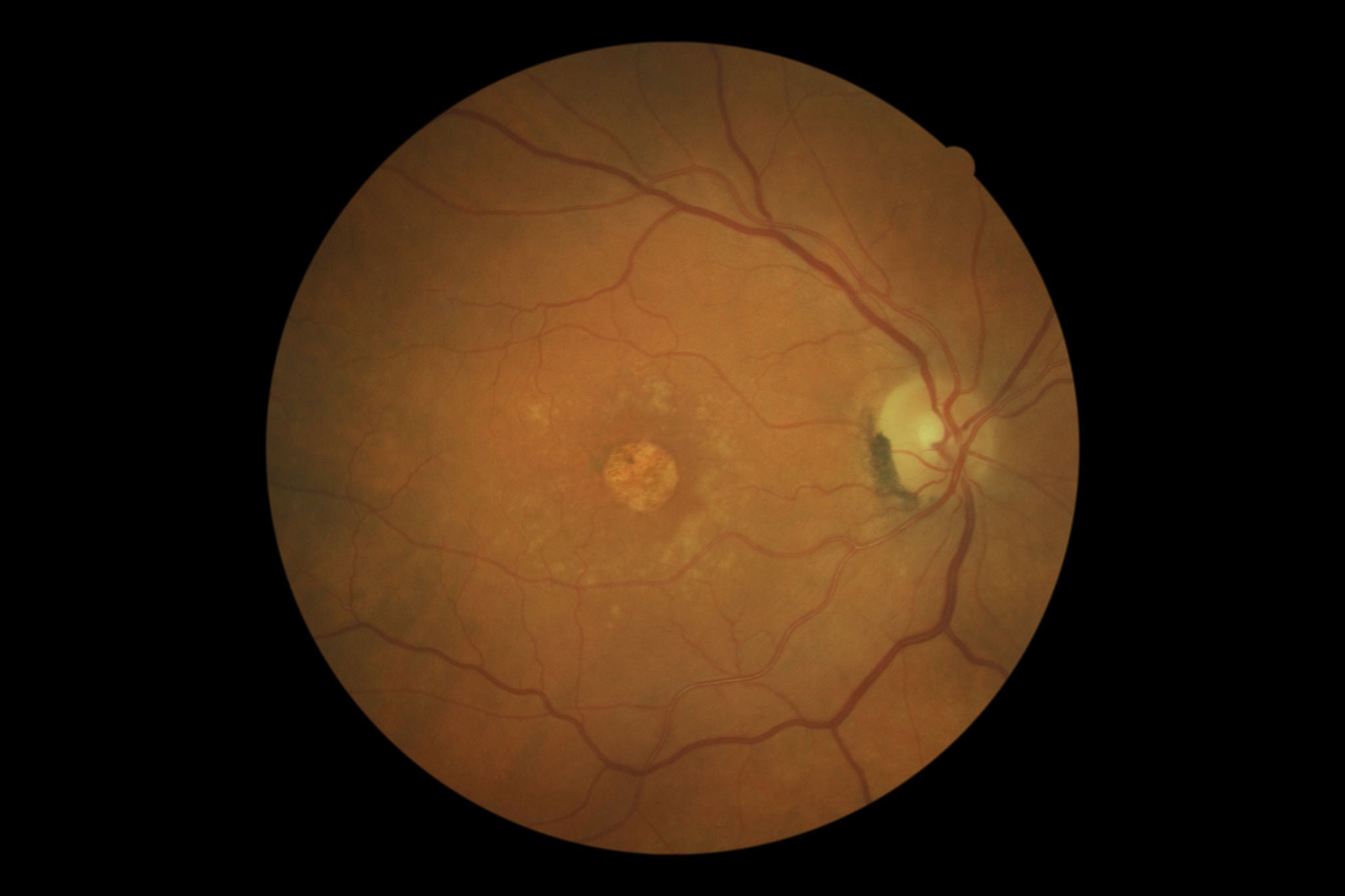
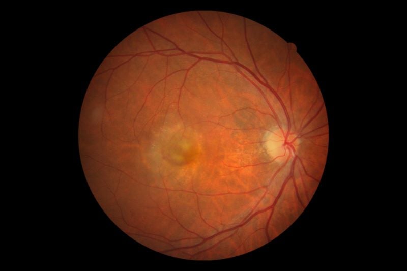 Color
Color
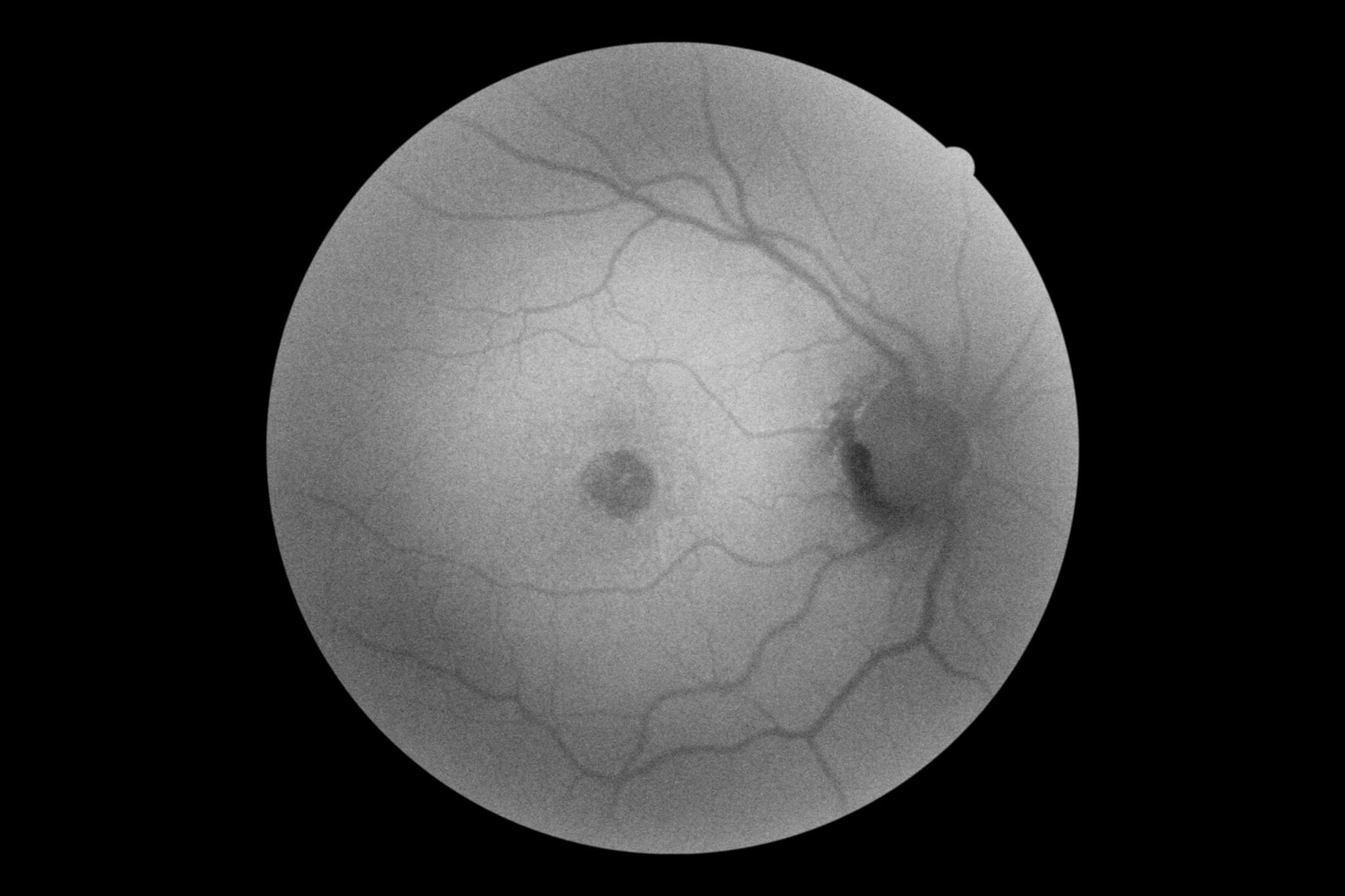
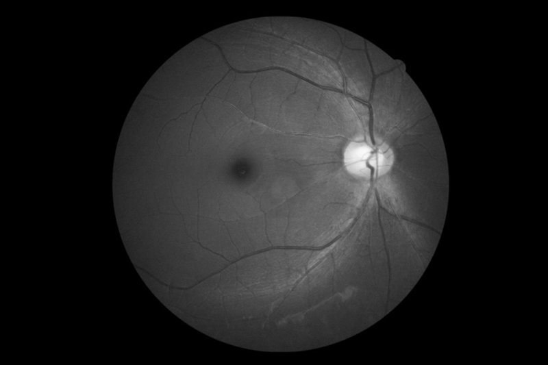 FAF
FAF
Low flash photography
In addition to the photometric Auto Exposure, the CR-2 Plus AF offers low flash photography. This feature greatly reduces the patients’ discomfort. Workflow is also improved as images of both eyes can be captured without being restricted by the pupils’ size. This is ideal for photophobic patients.
Fixation Lamp shifting presets
The CR-2 Plus AF offers the possibility to pre-set four fixation lamps patterns for internal fixation. Each pattern has a maximum of nine positions. This is an excellent feature when various screening protocols are used.
45 degrees
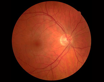
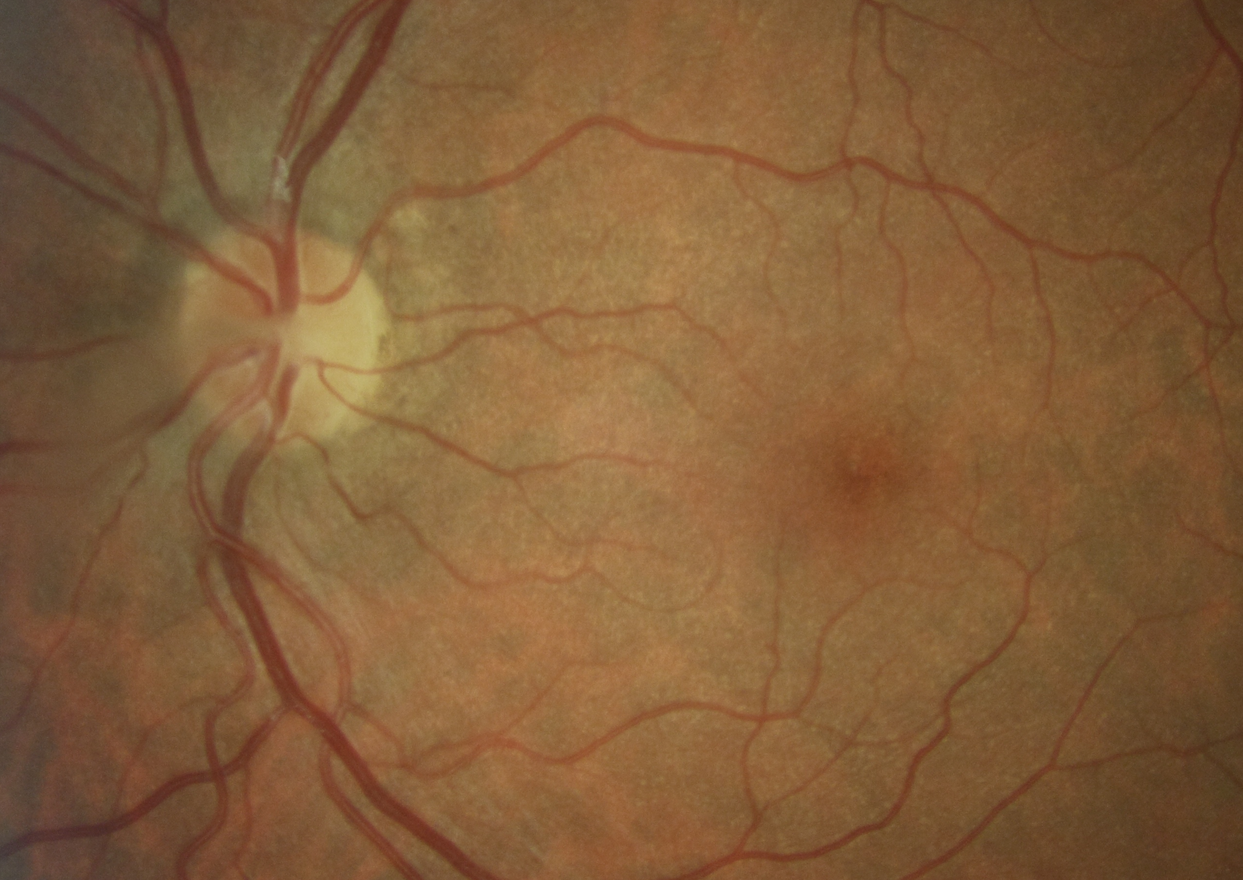
CR-2 Plus AF provides Non-Mydriatic images at a 45 degrees angle (67.5 degrees when using center of eye ball as reference).
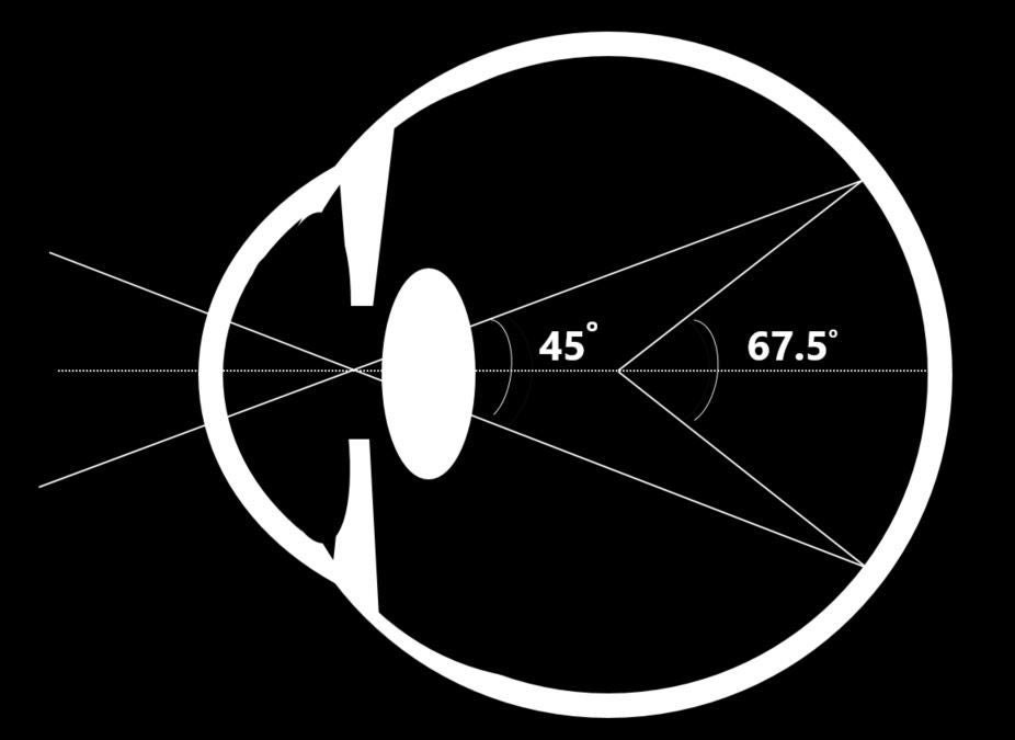 Angle of incidence vs central angle of view
Fundus Autofluorescence (FAF)
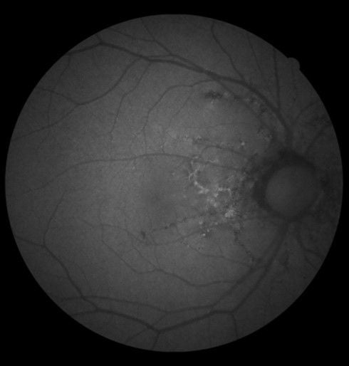
Fundus Autofluorescence (FAF) is a diagnostic technique for documenting the deposition of lipofuscin in the retinal pigment epithelium (RPE). As an easy and non invasive technique, it provides information that may otherwise not be clinically detectable. It can be a valuable asset in diagnosing retinal disease.
Since the introduction of fluorescein angiography (FFA) in 1959, ophthalmologists observed that even without the use of fluorescein, parts of the fundus would show areas of faint fluorescence under certain conditions. This naturally occurring fluorescence is mainly caused by Lipofuscin, a fluorescent pigment that accumulates in the RPE as a metabolic byproduct of cell function.
Lipofuscin deposition normally increases with age, but may also occur from RPE cell dysfunction or an abnormal metabolic load on the RPE. FAF imaging can visualize the deposition of lipofuscin in the retinal pigment epithelium (RPE). Areas of excess Lipofuscin accumulation will appear hyperfluorescent. However, when RPE cells die or are absent, LF disappears, leading to hypofluorescence.
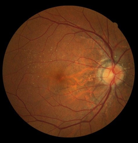
Am I missing something without FAF?
In this example, it is clear that the FAF image is showing additional clinical information that can’t be seen in the colour image!
Green Spectrum
The Canon CR-2 Plus AF utilizes green light spectrum to stimulate the retina and capture the emission in the yellow-orange spectrum while utilizing carefully selected matching optical filters. The green spectrum may provide more detail in the fovea, where as blue light tends to be absorbed by the high concentration of macular pigments. An additional advantage of green light is that the longer wavelength tends to have less absorption by the crystalline lens of the eye, especially in patients with cataracts.
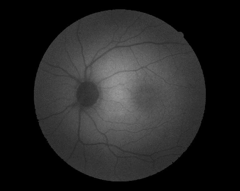
CLINICAL FAF images
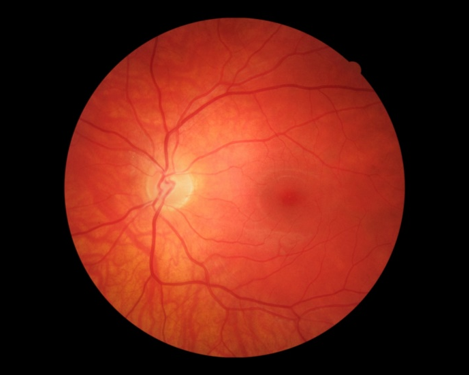
Normal distribution of lipofuscin
Retinal pigment epithelial atrophy

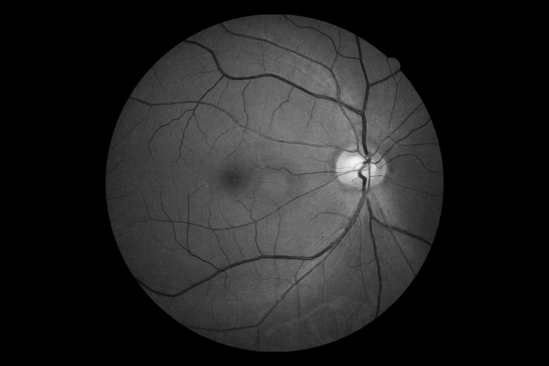
Digital Redfree and cobalt image
Without having the requirement to take an additional image, the RX software can create a digital red-free or cobalt image from a colour image. This is based on Canon’s EOS retina technology and its proprietary image processing that uses the digital camera’s original RAW image.
The image quality is fully comparable with those obtained via optical filters.
 Digital Redfree
Digital Cobalt
Digital Redfree
Digital Cobalt
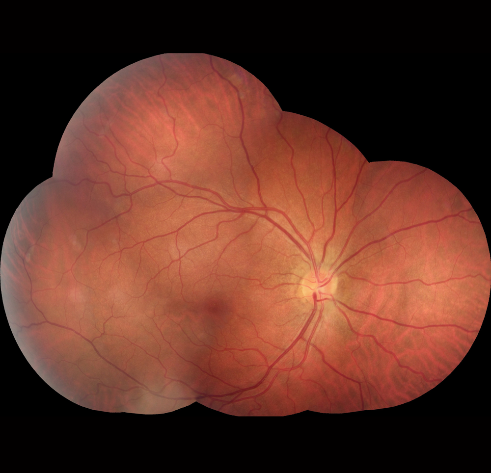
Stereo Photography
 Stereo guide marks
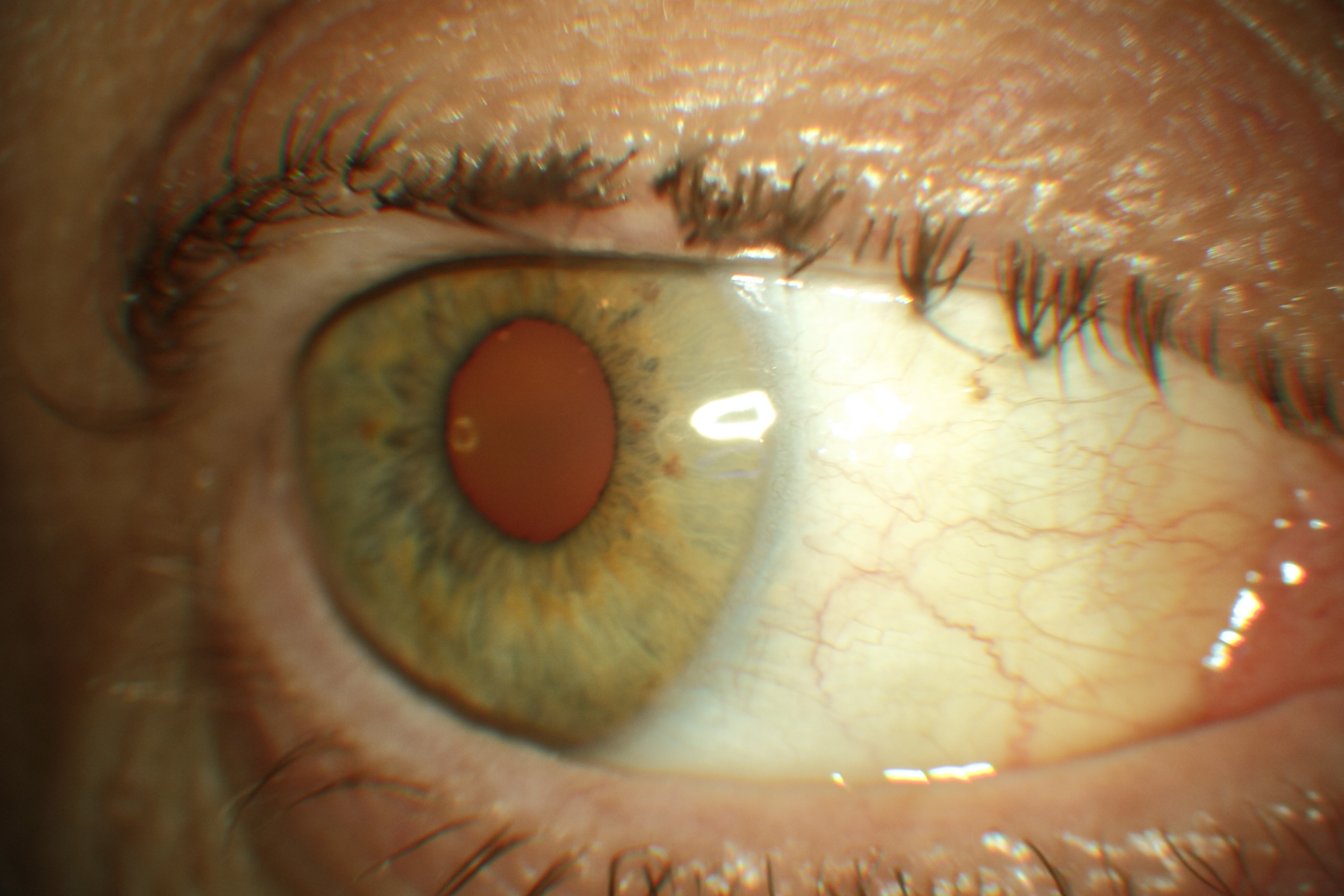
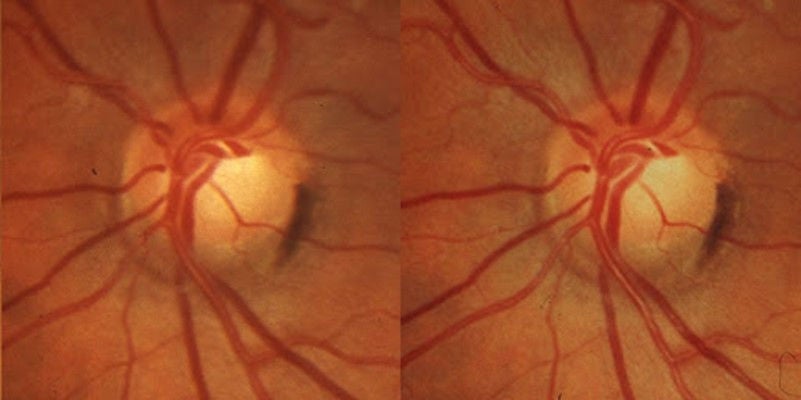
The CR-2 AF is suitable for stereo photography by sequentially taking two retinal images to form a stereo pair.
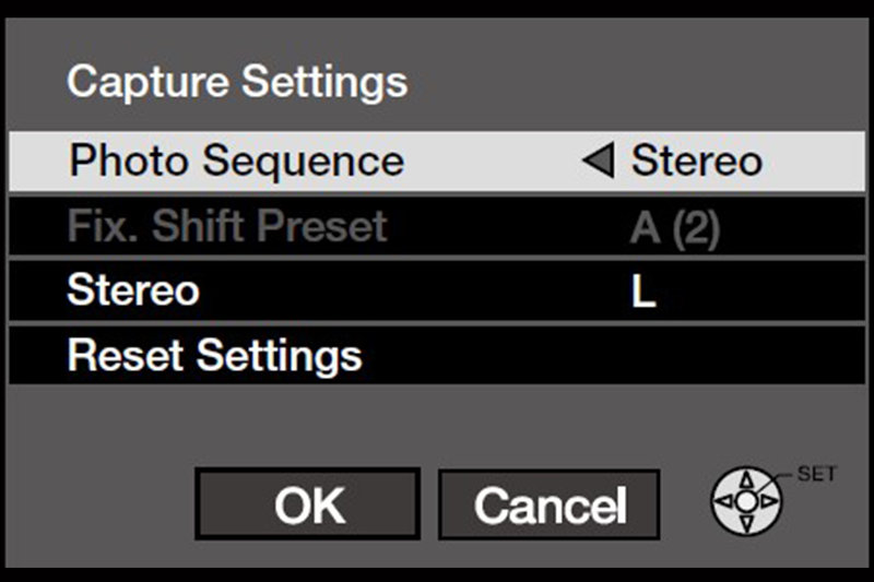
The capture sequence is simple to use with its stereo guides shown on the observation monitor. Alternatively, users can manually create a stereo set as well as a pair in the RX software.
Stereo viewing
 Stereo Pair
A standard commercial optical stereo viewer can be used for reviewing. The individual images of the stereo pair can easily be aligned in the RX software to obtain maximum stereo effect.
Social Media


