


Photon Counting CT
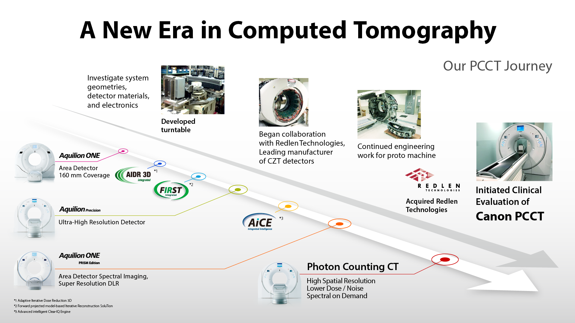
Since its inception in 1971, the dramatic evolution of Computed Tomography (CT) has led to the steady growth of CT’s role in patient care.
Canon Medical has pioneered many of the technological innovations defining the clinical expansion of CT, such as Area Detectors, Ultra-High Resolution, and Deep Learning Reconstruction. In partnership with Redlen Technologies Inc. (Redlen), a Canon group company, the global leader in photon counting detector design and manufacturing, Canon is currently developing a Photon Counting CT (PCCT) with the potential to improve visualization of small structures and enhance tissue characterization.
Our vision is to use Redlen’s state-of-the-art Photon Counting CT technology empowered by Canon Medical’s leading advancements in system, software, and image reconstruction to improve the quality of medical diagnosis for all patients worldwide.
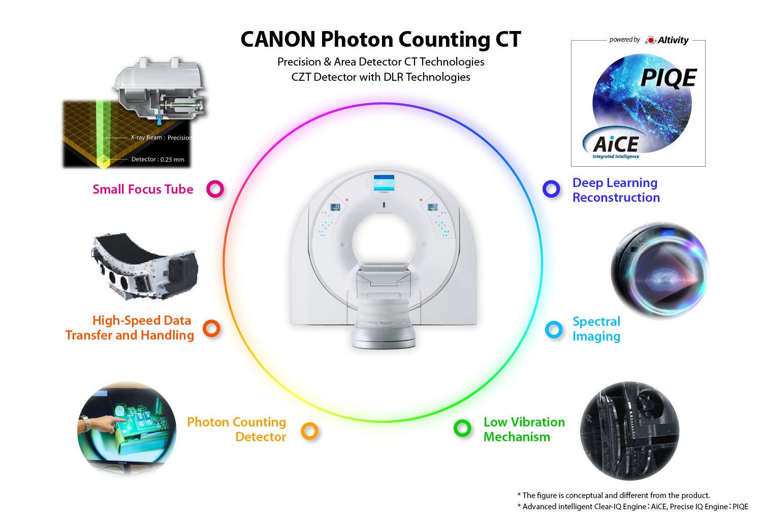
CANON'S PHOTON COUNTING CT
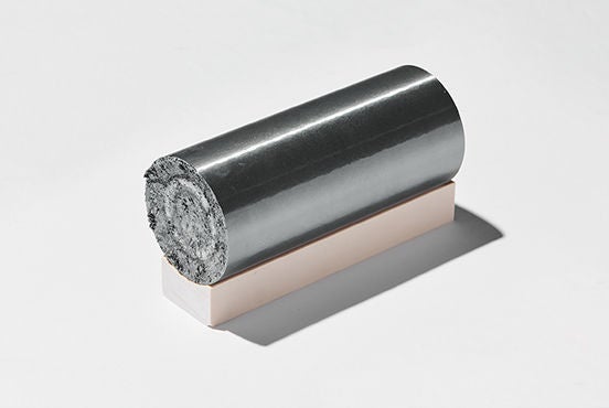
WHAT IS PHOTON COUNTING CT?
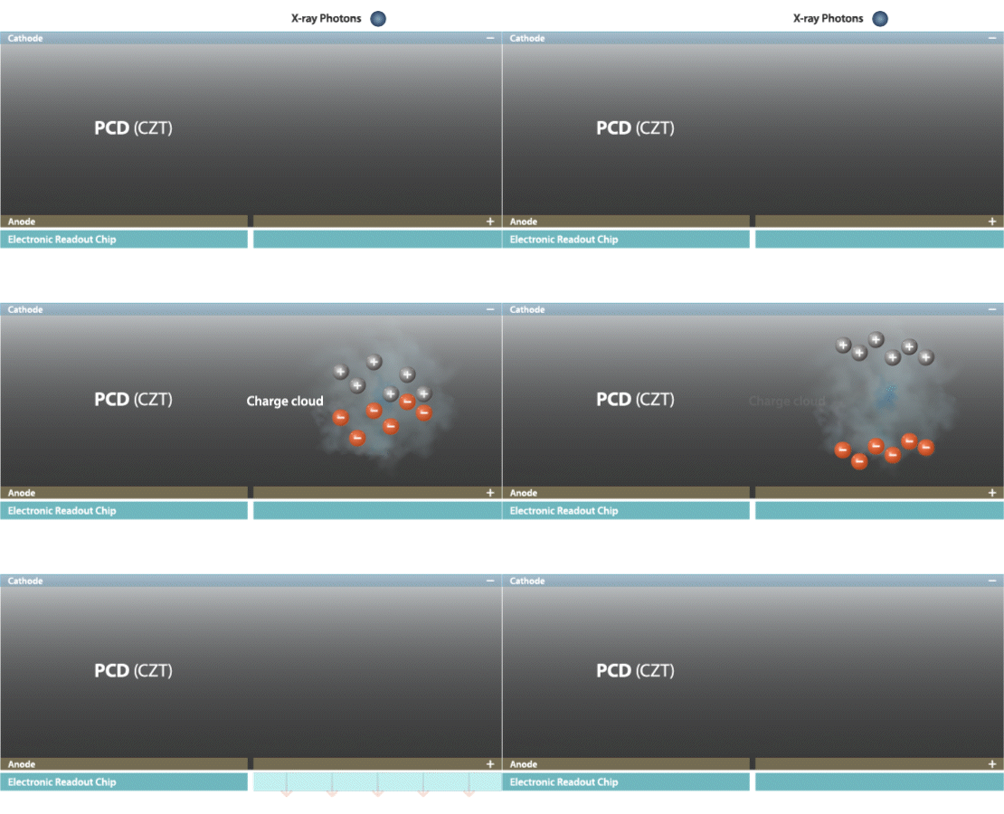
WHAT IS THE MAIN DIFFERENCE OF PHOTON COUNTING FROM CONVENTIONAL CT?
BENEFITS OF CANON’S PCCT: IMAGE QUALITY
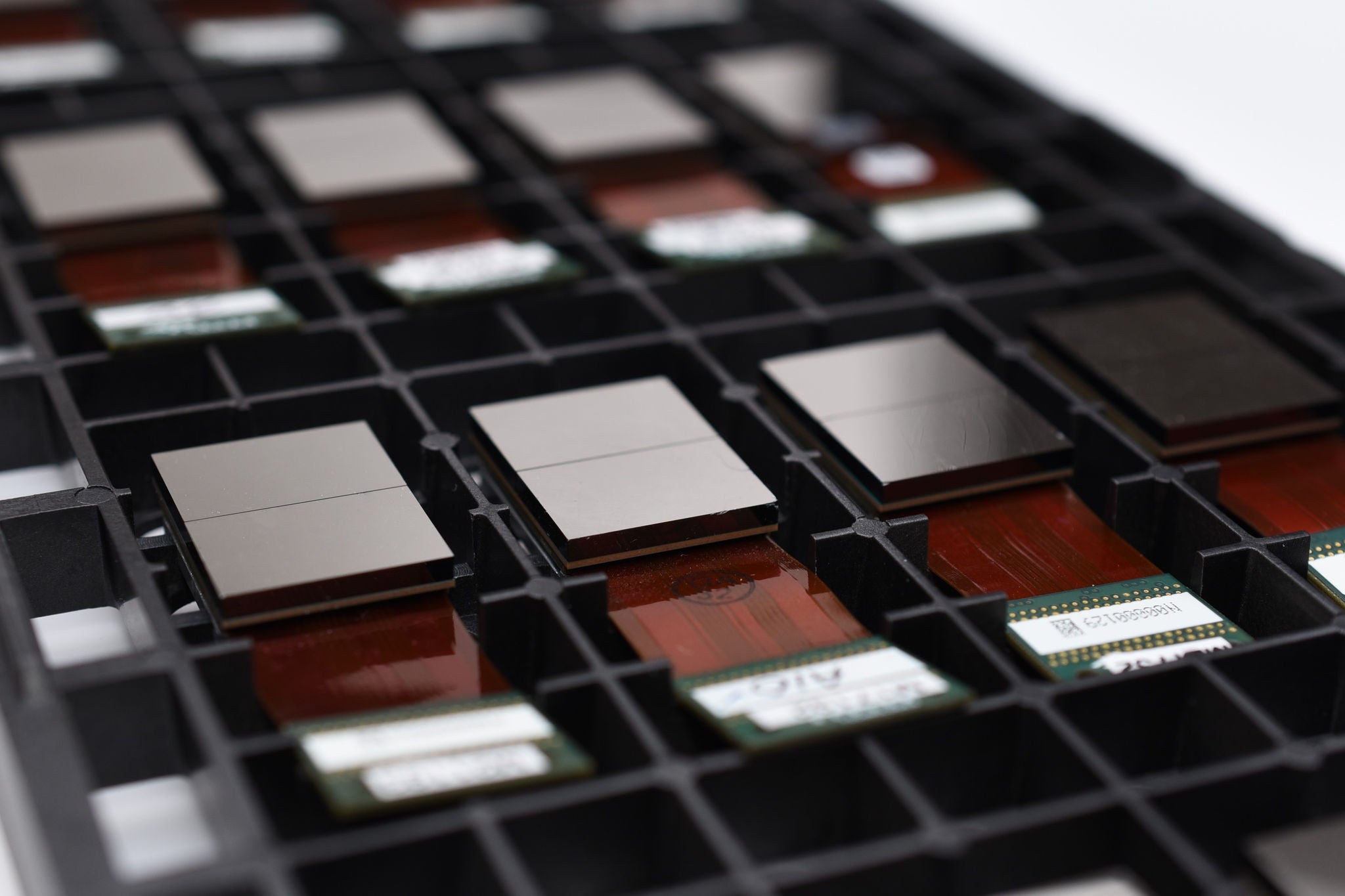
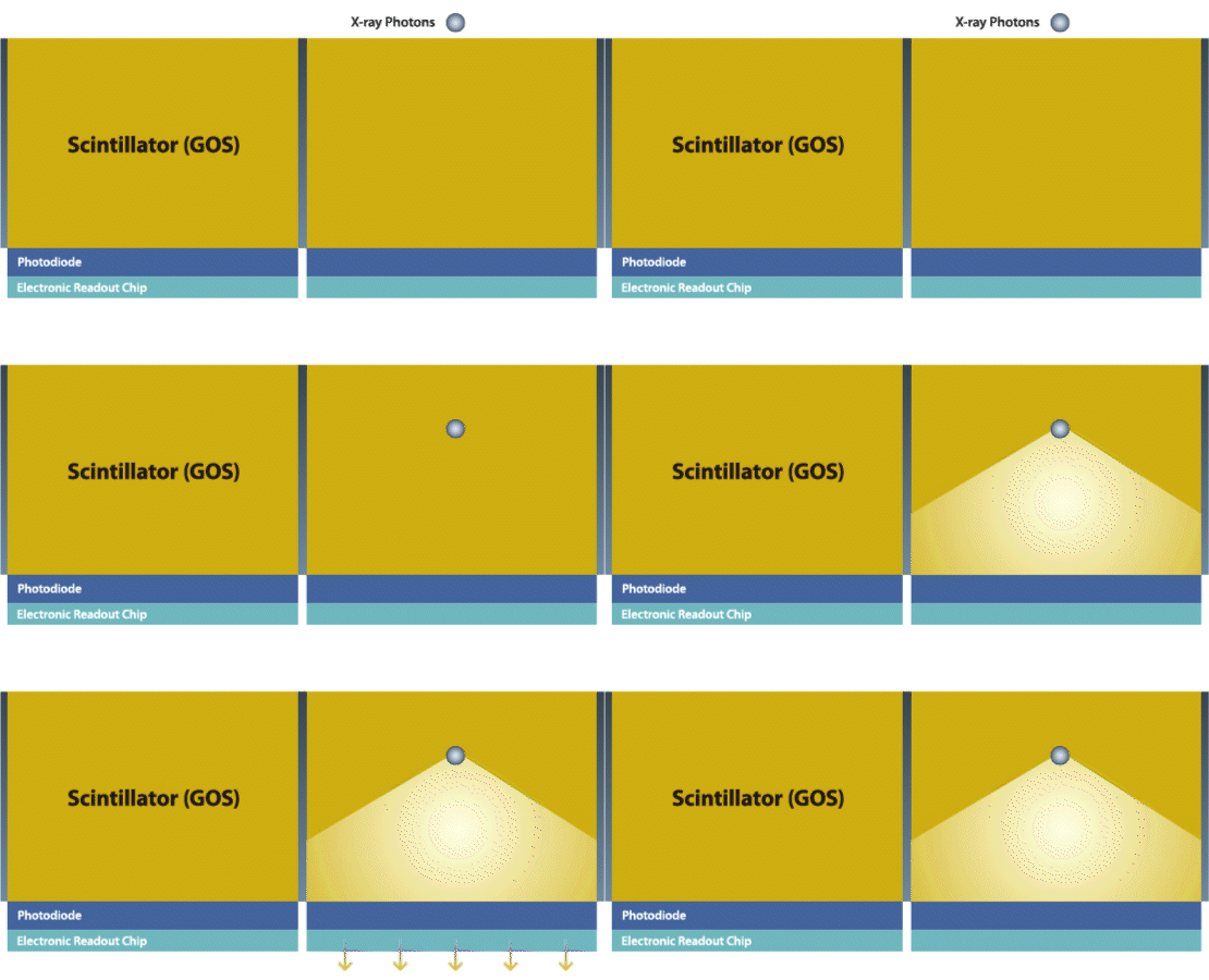
For more optimal dose efficiency, detectors should have as much active area capturing photons as possible. In conventional EIDs, light in one detector pixel can scatter into a neighboring pixel, a phenomenon called optical crosstalk that reduces spatial resolution. Because of this, EIDs require a reflector of finite thickness between the scintillator pixels to prevent crosstalk. However, the presence of this reflector reduces the active area of the detector and, thus, its dose efficiency, especially for small-sized detector pixels. Because PCCT doesn’t use a scintillator, there is no need for reflective material between detector pixels. This greatly improves the dose efficiency of the detector, allowing for smaller detector pixel sizes without dose penalty.
PCCT also overcomes a major disadvantage of EID: electronic noise. An EID’s electronic noise is unavoidably combined with true signal into the detector output. When the number of photons is low, electronic noise becomes dominant, degrading image quality. With PCCT, electronic noise from the detector registers below the threshold of lowest energy bin and is thus discarded. In this way, PCCT effectively eliminates electronic noise, which improves image quality.
BENEFITS OF CANON’S PCCT: SPECTRAL
PCCT enables Spectral Imaging with every scan for routine material decomposition. Canon’s exclusive advances in Spectral reconstruction have given Canon unique insight into minimizing noise and maximizing spectral information from PCCT. Because the thresholds for the energy bins are configurable, PCCT can also allow for imaging that targets specific K-edge energies, from common contrast agents, such as iodine and gadolinium, to novel nanoparticles such as gold.
BENEFITS OF CANON'S PCCT: ULTRA-HIGH RESOLUTION (UHR)
THE ADVANTAGE OF REDLEN, A CANON GROUP COMPANY


We're currently accumulating knowledge regarding both the technical and clinical benefits of our photon counting CT system.
Scientific papers
- Zhan X, Zhang R, Niu X, Hein I, Budden B, Wu S, Markov N, Clarke C, Qiang Y, Taguchi H, Nomura K, Muramatsu Y, Yu Z, Kobayashi T, Thompson R, Miyazaki H, Nakai H. Comprehensive evaluations of a prototype full field-of-view photon counting CT system through phantom studies. Phys Med Biol. 2023 Aug 14;68(17). doi: 10.1088/1361-6560/acebb3. PMID: 37506710.
- Lee D, Zhan X, Tai WY, Zbijewski W, Taguchi K. Improving model-data mismatch for photon-counting detector model using global and local model parameters. Med Phys. 2023 Dec 8. doi: 10.1002/mp.16883. Epub ahead of print. PMID: 38064641.
- Sasaki T, Kuno H, Nomura K, Muramatsu Y, Aokage K, Samejima J, Taki T, Goto E, Wakabayashi M, Furuya H, Taguchi H, Kobayashi T.CZT-based photon-counting-detector CT with deep-learning reconstruction: image quality and diagnostic confidence for lung tumor assessment.Jpn J Radiol. 2025 Mar 7. doi: 10.1007/s11604-025-01759-9. Online ahead of print. PMID: 40053285.
Conference presentations
- Y. Nakamura et al. Utility of CZT-based photon counting detector CT for a abdominal thin-slice non-contrast CT images in comparison with energy integrating detector CT. ECR 2025
- Y. Nakamura et al. Utility of virtual non-contrast images derived from CZT-based photon counting detector CT in comparison with tru non-contrast images. ECR 2025
- H. Kuno et al. Imaging-detected Extranodal Extension in Head and Neck Cancer: Clinical Implications and Diagnostic Criteria in the Era of High-Resolution Imaging including Photon-Counting Detector CT. RSNA 2024
- T. Sataki et al. CZT-based Photon-Counting-Detector CT with Deep-Learning Reconstruction: Image Quality and Diagnostic Confidence for Lung Tumor Assessment. RSNA 2024
- K. Nomura et al. Sharpness Evaluation of Chest Multi Planar Reconstruction Images with Normal and Super High-resolution Mode of CZT-Based Photon-counting Detector CT. RSNA 2024
- A. Pourmorteza et al. Dose-Efficient Characterization of Coronary Artery Plaques with a Prototype CdZnTe-Based Photon-Counting CT Scanner. SPIE 2024
- A. Pourmorteza et al. Iodine Quantification with a CdZnTe Clinical Prototype Photon-Counting Scanner At Reduced Radiation Dose: Initial Cardiac Phantom Results, ECR 2024
- K. Mei et al. Ultra-low-dose photon-counting CT: Assessing radiomic features with a patient-based lung phantom, ECR 2024
- S. Mochinaga et al. First Results of Electron Density Quantification with CZT-based Photon Counting Detector CT, ECR 2024
- W. Fukumoto et al. Comparison of newly developed CZT-based Photon Counting Detector CT (PCD-CT) and Ultra-High-Resolution CT (U-HRCT) for measuring airway dimensions: A phantom study. ECR 2024
- K. Yokomachi et al. Physical characteristics in slice direction using a newly developed CZT-based Photon-Counting Detector CT. ECR 2024
- Y. Nakamura et al. Accuracy of CT values on virtual monochromatic images of CZT-based Photon Counting Detector CT: comparison with dual-energy CT using energy integrating detector in a phantom model. ECR 2024
- T. Higaki et al. Utility of multi-energy mode of CZT-based Photon Counting Detector CT for coronary CT angiography: A structured phantom study. ECR 2024
- D. Lee et al. Advanced Photon-Counting Detector Simulator with a Count-Rate-Dependent Mapping Operator and a Pixel-to-Pixel Variation Generator. IEEE MSS MIC 2023
- A. Pourmorteza et al. Dose-efficient Ultra-high-resolution imaging of coronary stents with a CdZnTe-based clinical prototype photon- counting scanner. RSNA 2023
- K. L. Boedeker et al. Technical Performance of Super Resolution Deep Learning Reconstruction Algorithm on a Wide Area, Conventional Energy-Integrating Detector vs and a Photon-Counting Computed Tomography System with Conventional Reconstruction Algorithms. RSNA 2023
- T. Sasaki et al. CT Imaging of Lung Cancer: Exploring the Clinical Potential of CZT-based Photon Counting Detector CT. RSNA 2023
- K. Hirayama et al. Super-high-resolution abdominal imaging using CZT based photon counting CT with deep learning reconstruction: quantitative study and first clinical impression. RSNA 2023
- K. Nomura et al. Super-high-resolution chest imaging using CZT-based photon counting CT: performance characterization and first clinical trial. RSNA 2023
- S. Mochinaga et al. High z-axis resolution imaging using CZT based photon counting CT: quantitative study and first clinical trial. RSNA 2023
- Kei Mei et al. Evaluation of a prototype photon-counting CT for pulmonary imaging using patient-based lung phantoms. RSNA 2023
- S. Kondo et al. Visualization of simulated small vessels on photon counting detector CT: comparison with energy integrating CT in a phantom model. RSNA 2023
- T. Higaki et al. Improving spatial resolution in coronary CT angiography with photon counting detector CT: A structured phantom study. RSNA 2023
- T. Higaki et al. Noise reduction in coronary CT angiography with photon counting detector CT: A structured phantom study. RSNA 2023
- F. Tatsugami et al. Coronary Artery Calcium Volume Measurement: A Comparison between Photon-Counting CT and Ultra-High-Resolution CT using a Cardiac CT Calibration Phantom. RSNA 2023
- K. Nomura et al. Basic Image Quality Evaluation of New Platform Prototype Photon Counting CT. JRC 2023
- X. Zhan,et al. Spectral imaging performance evaluation for a prototype full-size photon counting CT system at clinical dose levels. JRC 2023
- R. Zhang,et al. Quantitative image quality comparison between normal resolution and super high resolution modes of a clinical prototype photon counting CT system. JRC 2023
- T. W. Holmes et al. Pediatric head and neck imaging with a CZT-based photon-counting CT scanner : initial image quality evaluation. ECR 2023
- K. Nomura et al. Comparison of CT image quality for different sized phantom between prototype full-size photon counting and conventional CT systems : CT number, image noise and artifact. ECR 2023
- Edgar Salazar et al. Evaluation of a prototype photon-counting CT for low-dose pulmonary imaging using patient-based lung phantom. ECR 2023
- X. Zhan et al. A study of cross-talk effect in pixelated photon counting detectors and impact to system imaging performance. SPIE 2023
- Donghyeon Lee et al. Photon-Counting Detector Model Using Local Parameters For Pixel-to-Pixel Variation. SPIE 2023
- W. Yang Tai et al. Effects of Bowtie Scatter on Material Decomposition in Photon-Counting CT. SPIE 2023
- R. Zhang et al. Quantitative Image Quality Comparison between Photon Counting and Conventional CT Systems: Contrast-to-Noise Ratio. RSNA 2022
- K. L. Boedeker et al. Low Contrast Detectability Comparison Between a Prototype Photon Counting Computed Tomography System and Conventional CT system Across a Range of Attenuation Levels. RSNA 2022
- Xiaohui Z et al. Quantitative image quality evaluation for a prototype photon counting CT through phantom studies: Noise, Resolution and Quantitative Accuracy. CERN SpecXray 2022
- A. Pourmorteza et al. First experience with a clinical prototype CZT-based PCCT scanner: applications in low-dose lung cancer screening. CERN SpecXray 2022
- Xiaohui Z et al. Phantom imaging evaluations of a prototype CZT based photon counting system. ECR 2022
- K. Nomura et al. Quantitative image quality comparison between a prototype full-size photon counting CT system and conventional CT systems with energy integrating detectors. ECR 2022
- T. W. Holmes et al. Low-Dose Lung Cancer Screening with a Novel CZT Photon-Counting CT Prototype: A Phantom Study. ECR 2022
- Y Suzuki et al. Physics Performance Evaluation of Prototype Photon Counting CT: Basic Image Quality Evaluation. JRC 2022
- K. Nomura et al. Physics Performance Evaluation of Prototype Photon Counting CT: Quantitative Evaluation. JRC 2022
- Y. Muramatsu et al. Physics Performance Evaluation of Prototype Photon Counting CT: Large-phantom Evaluation. JRC 2022
- Xiaohui Z et al. First results from a prototype full-size photon counting CT system: counting and spectral imaging performance at clinical dose levels. RSNA 2021
- K. Nomura et al. Quantitative image quality comparison between photon counting and conventional CT systems: noise, resolution and quantitative accuracy. RSNA 2021
Press Releases
August 31, 2021
Commencement of Research Collaboration into Japan's first Photon Counting Computed Tomography with National Cancer Center Japan (medical.canon)
November 7, 2022
First Domestically Produced X-ray CT System with Photon-counting Detector Installed at National Cancer Center Japan Exploratory Oncology Research & Clinical Trial Center (medical.canon)
April 12, 2023
Start of Clinical Research of the First Japanese Produced Next-Generation Photon-Counting Computed Tomography
November 17, 2023
Canon Medical Systems Accelerates Global Clinical Research to Realize Next-Generation Photon-Counting CT
February 26, 2024
Clinical Research on Photon-Counting CT Begins with Radboud University Medical Center
April 8, 2024
Start of Clinical Research with Hiroshima University on Photon-Counting CT
November 27, 2024
Canon Launches Research Collaboration with Penn Medicine for Application of Photon-Counting CT
Disclaimer
Note: Photon Counting CT technology is currently under development and the subject of ongoing research and development. Each technology is not yet commercialized and is not available for sale.
Social Media


